Electrophoresis and Its application in Pharmacognosy
Electrophoresis is a laboratory technique used to separate and identify proteins, enzymes, amino acids, nucleic acids, and other biomolecules present in tissues, based on their size and electrical charge.
An electric current is used to move the molecules through a gel or other matrix. Pores in the gel or matrix work like a sieve, allowing smaller molecules to move faster than larger molecules. To determine the size of unknown molecules in a sample, standard compounds of known molecular sizes are run on the same gel under the same electrophoretic conditions. The migration of the unknown molecules is then compared with that of the standards to estimate their molecular size and, in some cases, help identify them. The rate of migration of an ionic compound in electrical field depend on some factors,- Net charge of molecule
- Size and shape of particle
- Strength of electrical field
- Properties of supporting medium and
- Temperature of operation
Basic Principle of Electrophoresis
Basic Principle of Electrophoresis
Electrophoresis is an analytical technique in which charged particles move towards the oppositely charged electrode under the influence of an applied electric field, resulting in their separation.
The movement of particles depends mainly on their net electrical charge, size, shape, buffer pH, and the properties of the supporting medium.- Cations (positively charged particles) migrate towards the cathode (−).
- Anions (negatively charged particles) migrate towards the anode (+).
- Particles with different electrophoretic mobilities migrate at different rates, allowing them to be separated.
- Particles with zero net charge generally show little or no electrophoretic migration under the same conditions.
| Type | Application |
|---|---|
| Paper electrophoresis | Separation of amino acids and other small charged molecules |
| Agarose gel electrophoresis | DNA and RNA analysis |
| PAGE (polyacrylamide gel electrophoresis) | Protein and smaller DNA fragments separation |
| SDS-PAGE | Separation of proteins mainly according to molecular mass specially from 5 to 250 kDa |
Paper Electrophoresis
Paper electrophoresis is a technique used to separate charged molecules on a strip of cellulose filter paper by applying an electric field. It is mainly used for the separation and analysis of amino acids, peptides, and other small charged biomolecules.
Paper Electrophoresis
Basic Principle of Paper Electrophoresis
Basic Principle of Paper Electrophoresis
Paper electrophoresis is a separation technique in which a strip of cellulose filter paper acts as the supporting medium. The paper is first soaked in a suitable electrophoresis buffer (running buffer) and placed between two buffer reservoirs connected to a power supply.
The sample is applied as a small spot near the centre or one end of the paper, and an electric field (typically 100–300 V, depending on the apparatus and length of the paper) is applied across it. Under the influence of the electric field, charged molecules migrate through the moist paper toward the electrode of opposite charge. The rate and direction of migration depend primarily on the net charge, size, and shape of the molecules, as well as the pH and ionic strength of the buffer. Molecules carrying a greater net charge migrate faster, whereas molecules with lower charge or larger size migrate more slowly. At their isoelectric point (pI), molecules possess no net charge and therefore show little or no migration. Since the pores of filter paper are relatively large, paper electrophoresis separates molecules mainly on the basis of their electrical charge rather than molecular size. It is most suitable for the separation and analysis of amino acids, peptides, small proteins, nucleotides, and other charged biomolecules, and has been widely used in biochemical and clinical laboratories.
Gel Electrophoresis
Gel electrophoresis is a laboratory technique used for the separation of charged biomolecules such as DNA, RNA, and proteins by passing them through a porous gel matrix under the influence of an electric field.
Gel electrophoresis is mainly classified based on the type of gel matrix and the separation method used. The two important types are Agarose Gel Electrophoresis and Polyacrylamide Gel Electrophoresis (PAGE). Agarose gel electrophoresis is commonly used for the separation of DNA and RNA fragments due to its relatively larger pore size. Polyacrylamide Gel Electrophoresis (PAGE) provides higher resolution and is mainly used for the separation of proteins and small nucleic acid fragments. A specialized form of PAGE, known as SDS-PAGE (Sodium Dodecyl Sulfate–Polyacrylamide Gel Electrophoresis), separates proteins primarily according to their molecular size (molecular weight) because SDS imparts a uniform negative charge to proteins, eliminating differences due to their native charge and shape.
Gel Electrophoresis
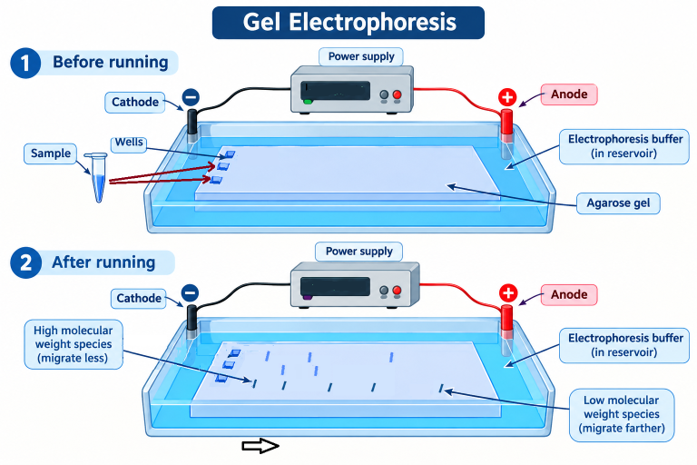
Basic Principle of Gel Electrophoresis
Basic Principle of Gel Electrophoresis
Gel electrophoresis is a separation technique in which a porous gel matrix acts as a molecular sieve. The gel, usually agarose or polyacrylamide, is immersed in a suitable electrophoresis buffer (running buffer), and an electric field is applied across the gel (typically 50–150 V, depending on the gel size, thickness, and experimental requirements).
Charged molecules migrate through the pores of the gel toward the electrode of opposite charge. Smaller molecules pass through the pores more easily and therefore migrate faster. Whereas larger molecules experience greater resistance within the gel matrix and migrate more slowly, covering a shorter distance in the same time. Thus, separation occurs primarily on the basis of molecular size, although the charge and shape of the molecules can also influence their migration. The choice of gel matrix depends on the type of molecules being analyzed. Agarose gel, having relatively large pores, is best suited for the separation of DNA and RNA fragments, especially larger nucleic acid molecules. In contrast, polyacrylamide gel (PAGE) has much smaller and more uniform pores, making it ideal for the high-resolution separation of proteins, peptides, and small DNA fragments. Therefore, selecting an appropriate gel matrix is essential for achieving efficient and accurate separation of biomolecules.| PAGE | SDS-PAGE | Proteins are separated according to a combination of their net charge, size, and shape while largely retaining their native structure. | Proteins are denatured using sodium dodecyl sulfate (SDS), which gives them a slight negative charge-to-mass ratio. They are separated mainly according to molecular mass. | Proteins generally retain their folded structure and may retain biological activity. Separation depends on charge and size. | Proteins are denatured and separated mainly according to molecular mass. Smaller proteins generally travel farther through the gel than larger proteins. | The proteins are stable in a Native PAGE, and can be recovered later. | The proteins are not stable in the SDS PAGE, and hence cannot be recovered. | Used to study the structure, subunit composition and function of proteins; purify proteins from a mixture in native form. | To detect presence of a protein; determine molecular weight (MW) of a protein; check protein expression etc. |
|---|